


What is IVF
In Vitro Fertilization is a highly effective treatment to help patients achieve pregnancy using their eggs and sperm

In Vitro Fertilization (IVF) is a highly effective treatment to help patients achieve pregnancy using their eggs and sperm.
We use gentle and individualized ovarian stimulation protocols prioritizing egg quality over quantity. This approach minimizes clinic visits and reduces the risk of ovarian hyperstimulation without compromising the chances of a successful outcome. Most patients can maintain their everyday lifestyles during treatment.
In Vitro Fertilization Procedure
In Vitro Fertilization procedure consists of the following steps:
- Ovarian stimulation
- Egg retrieval procedure
- Fertilization of eggs
- Culture of embryos
- Assisted hatching of embryos (when indicated)
- Embryo transfer
- Implantation
1. Ovarian Stimulation
There are various forms of ovarian stimulation protocols, each with multiple modifications. Your treatment is always personalized to maximize the likelihood of success. The selection of the optimal protocol for your IVF treatment is based on your reproductive history and your pre-treatment evaluation.
Below is an example of an IVF treatment cycle. Your individualized protocol may take less or more time to complete.
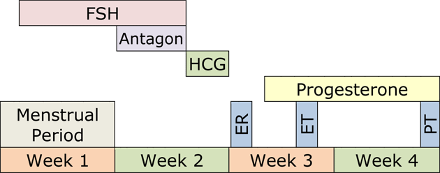
Follicle stimulating hormone (FSH), or a combination of FSH and luteinizing hormone (LH), is used to stimulate the production of multiple eggs in the ovaries. These hormones are administered subcutaneously once a day or every other day using tiny needles for approximately ten days.
Throughout this period, your progress is monitored through estradiol (estrogen, E2), progesterone blood levels, and ultrasound examinations.
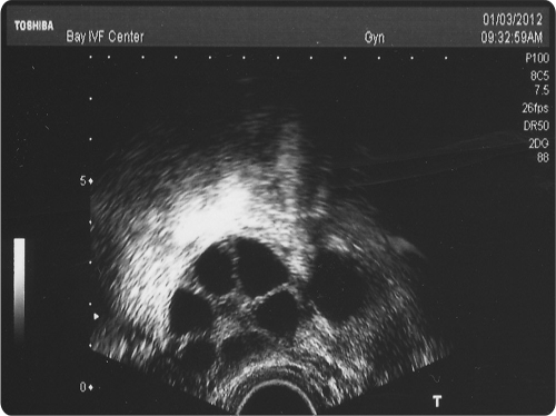
Ovarian stimulation should result in the development of several eggs in each ovary. The ultrasound image below shows a stimulated ovary. Each of the several follicles (dark circles) contains a microscopic egg.
2. Egg Retrieval Procedure
The egg retrieval procedure is performed at our clinic. It only takes a few minutes, and we provide comfortable conscious sedation for analgesia.
Under ultrasound guidance, a thin needle is inserted through the top of the vagina into the cul-de-sac (space behind the uterus). The ovaries are located near the bottom of the cul-de-sac allowing the needle to access the ovarian follicles and aspirate the follicular fluid. The fluid is examined under a microscope to identify the eggs.
3. Fertilization of Eggs
On average, eight to fourteen eggs are retrieved during the egg retrieval procedure. Once identified, the eggs are placed in petri dishes filled with culture medium. The composition of the culture medium resembles the fluid secreted by the Fallopian tubes, facilitating the development of eggs and embryos (fertilized eggs) in our laboratory environment, simulating the conditions in the Fallopian tubes.
On the day of egg retrieval, the male partner provides a semen specimen through masturbation. The highest quality sperm are extracted from the semen and combined with the eggs three hours after retrieval. This in vitro fertilization process takes place over several hours in the evening following egg retrieval.
If the male partner has never caused pregnancy or if his test results indicate significant male infertility, Intracytoplasmic Sperm Injection (ICSI) is performed. In ICSI, a single sperm is directly injected into an egg, significantly increasing the probability of successful fertilization for selected patients.
4. Culture of Embryos
Fertilization is usually observed the day after insemination, approximately 16 hours later. A normally fertilized egg (zygote) will exhibit two pronuclei representing the genetic material from the egg and sperm. The fertilized eggs are transferred to a growth medium and continue to be cultured in our laboratory.
The following day, embryos should divide into 4 cells, and by the day after, they should reach 8 cells.

By the fifth to seventh day after the insemination, the embryos should develop into the blastocyst stage, consisting of 80 or more cells.
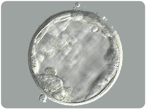
The picture below shows an advanced stage of blastocyst development. Notice the central fluid-filled cavity. The cells within the blastocyst have already differentiated into the inner cell mass (at seven o’clock) that will give rise to the fetus and the trophectoderm cells that will form the future placenta.
At this stage, the embryos are still microscopic, invisible to the naked eye.
5. Assisted Hatching
Assisted hatching is a laboratory procedure used to create a “weak spot” in the eggshell of an embryo. This technique is primarily employed for cryopreserved embryos, as it increases the likelihood of successful hatching, implantation, and, ultimately, a healthy baby.
This picture shows an embryo after assisted embryo hatching with an opening breaching the eggshell at the 12 o’clock position.

6. Embryo Transfer
The embryo transfer procedure typically occurs five days after egg retrieval, depending on the development of the embryos. Prior to the transfer, our embryologists assess the embryos to determine their chances of successful implantation. This assessment helps you decide whether to transfer one or two embryos.
Just before the embryo transfer, the embryo(s) is/are placed into the tip of a thin embryo transfer catheter. The catheter is then passed through the cervical canal, reaching within 15 mm of the top of the uterine cavity, where the embryos are gently released.
The embryo transfer itself usually takes only a few seconds to complete, and no rest is required afterward.
In cases where there are more embryos than the intended number for transfer, these embryos can be frozen and stored in liquid nitrogen. The implantation success rate of frozen embryos is equal to that of “fresh” embryos.
7. Implantation
After the embryo transfer, the endometrial lining gently holds the embryo(s) within the upper part of the uterus. There are no restrictions on your physical activity during this period.

This picture shows a healthy blastocyst in the process of “squeezing out” of its eggshell. Once fully hatched, it will stay in the uterus unattached for one to two days and then implant.
The lining of the uterus becomes receptive to the embryos through the action of estrogen and progesterone hormones produced by the ovaries. Ovarian progesterone production is supplemented with vaginal progesterone capsules.
A blood pregnancy test is scheduled two weeks after the embryo transfer. If the test is positive, an ultrasound examination is scheduled two weeks later to visualize the implantation site and check for a heartbeat within the embryo. Once a heartbeat is detected, there is a 95% probability that the pregnancy will result in a live birth.

This ultrasound picture shows a six-week pregnancy. The pregnancy sac is 25 mm in diameter. The baby inside the sac is only 13 mm long. It is already possible to distinguish the baby’s head and body and to see the cardiac activity.
At this point, the pregnancy becomes indistinguishable from natural conception, and your obstetrical care should proceed as it would if you conceived without any treatment.


Schedule Your Initial Consultation With Dr. Polansky
Online (no cost) or In-Person
Call or Text Us: Call or text us at 📞 650 322 0500
You can also complete the form below to request your initial consultation.


Let’s talk about your next step